Implant assessment
Assessment of available bone and its relationship to nearby anatomical structures.
Explore teeth, jaws and their spatial relationships in three dimensions, when additional information is needed for your care.
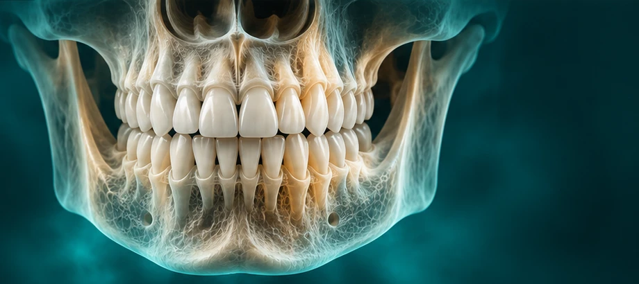 3D VOLUME · ILLUSTRATIVE VISUAL
Cone beam computed tomography uses X-rays to create a three-dimensional view of dental and bony structures. Your dentist selects this examination when it can answer a clinical question that simpler imaging cannot adequately resolve.
Your referring clinician determines whether this examination is appropriate.
Assessment of available bone and its relationship to nearby anatomical structures.
Additional information in specific complex cases, following clinical assessment.
Three-dimensional localisation when clinically indicated.
Cross-sectional information about teeth and bony anatomy, reviewed in axial, coronal and sagittal planes. A rendered 3D view helps orientation; assessment relies on the underlying volume.
CBCT has limited soft-tissue contrast. Motion and dense dental materials can obscure detail. Findings need to be interpreted with the referral and clinical assessment.
A volume for selected spatial or cross-sectional questions.
A panoramic 2D overview of the teeth and jaws.
A side-view 2D radiograph used in orthodontic assessment.
Include the question you need answered, rather than the modality alone. The acquisition and reporting requirements should be agreed for the individual examination.
Explore the referral pathwayBring your dentist’s prescription and any relevant earlier images. Let the team know about pregnancy or possible pregnancy and any positioning or mobility needs.
Read the patient guideThe requested examination and area should be clear.
The team will explain the examination before acquisition.
Discuss the relevant image and report arrangements.
Explore common imaging applications for 3D CBCT.
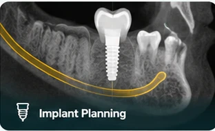 Illustrative visualAnatomy & spatial relationshipsImplant Planning
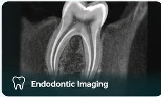 Illustrative visualSelected tooth-related questionsEndodontic Imaging
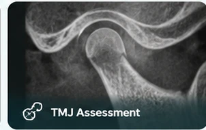 Illustrative visualBony joint anatomyTMJ Assessment
Illustrative visuals only. These are not DentoRad patient scans, findings or reports.
No. OPG is a two-dimensional panoramic image; CBCT creates three-dimensional information. The right examination depends on what your dentist needs to assess.
DentoRad offers specialist CBCT reporting. The reporting scope and delivery arrangements should be confirmed for your examination.
The area and protocol should follow the referral and clinical assessment. Bring the prescription and any relevant earlier images.
A two-dimensional overview of the teeth and jaws, supporting your dentist’s assessment and treatment planning.
Explore Digital OPGA prescribed side-view dental radiograph to support assessment of the relationship between the teeth, jaws and facial profile.
Explore Lat Ceph2D & 3D TMJ EXAMINATIONSTwo-dimensional and three-dimensional imaging options for selected jaw-joint questions, with a clear understanding of what each can show.
Explore TMJ ImagingExplore your examination or arrange a dental imaging visit.