Orthodontic records
A lateral view as part of a clinically indicated orthodontic assessment.
A prescribed side-view dental radiograph to support assessment of the relationship between the teeth, jaws and facial profile.
A lateral cephalogram creates a side-view image of the head. Your orthodontist may use it alongside examination findings and other records when planning treatment.
Your referring clinician determines whether this examination is appropriate.
A lateral view as part of a clinically indicated orthodontic assessment.
Information about the relationship of the upper and lower jaws.
Comparison with earlier records when the treating clinician considers it necessary.
A reproducibly positioned lateral projection that supports assessment of skeletal and dental relationships. The orthodontist considers it with clinical findings and other orthodontic records.
A lateral image compresses 3D anatomy into a 2D projection with overlapping right and left structures. It does not provide the same information as a CBCT volume.
Obtaining the prescribed lateral radiograph.
Landmark identification and measurements; confirm the requested scope.
The orthodontist’s clinical decision using all relevant records.
Include the question you need answered, rather than the modality alone. The acquisition and reporting requirements should be agreed for the individual examination.
Explore the referral pathwayBring your dentist’s prescription and any relevant earlier images. Let the team know about pregnancy or possible pregnancy and any positioning or mobility needs.
Read the patient guideThe requested examination and area should be clear.
The team will explain the examination before acquisition.
Discuss the relevant image and report arrangements.
Yes. Lat Ceph is a common shortened name for a lateral cephalogram.
No. A lateral cephalogram is a side view; an OPG is a panoramic view of the teeth and jaws.
Your orthodontist combines the image with clinical findings and other records to make treatment decisions.
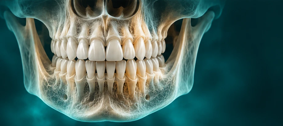 THREE-DIMENSIONAL IMAGING
Explore teeth, jaws and their spatial relationships in three dimensions, when additional information is needed for your care.
Explore 3D CBCTA two-dimensional overview of the teeth and jaws, supporting your dentist’s assessment and treatment planning.
Explore Digital OPG 2D & 3D TMJ EXAMINATIONS
Two-dimensional and three-dimensional imaging options for selected jaw-joint questions, with a clear understanding of what each can show.
Explore TMJ ImagingExplore your examination or arrange a dental imaging visit.