TMJ X-ray
Two-dimensional imaging for selected questions, with known limitations from overlap and projection.
Two-dimensional and three-dimensional imaging options for selected jaw-joint questions, with a clear understanding of what each can show.
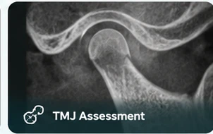 TMJ ANATOMY · ILLUSTRATIVE VISUAL
The temporomandibular joints connect the lower jaw to the skull. Imaging is selected after clinical assessment. CBCT can help assess bony joint structures, while other methods may be needed for soft tissues.
Your referring clinician determines whether this examination is appropriate.
Two-dimensional imaging for selected questions, with known limitations from overlap and projection.
Cross-sectional assessment of bony joint anatomy when clinically justified.
CBCT does not adequately show the joint disc. MRI may be required for suspected disc or other soft-tissue problems.
The requested modality is chosen for the suspected problem. CBCT provides bony joint detail; 2D projection images provide a more limited perspective.
Neither a 2D TMJ X-ray nor CBCT adequately evaluates the articular disc. MRI may be needed for disc position or other soft-tissue questions. MRI is not presented as a DentoRad service.
2D projection views; superimposition limits assessment.
Cross-sectional evaluation of bony joint anatomy.
A different modality for disc and soft-tissue assessment.
Include the question you need answered, rather than the modality alone. The acquisition and reporting requirements should be agreed for the individual examination.
Explore the referral pathwayCross-sectional views of the bony joint structures.
Questions about the disc may require MRI. MRI is discussed here for context and is not listed as a DentoRad service.
Bring your dentist’s prescription and any relevant earlier images. Let the team know about pregnancy or possible pregnancy and any positioning or mobility needs.
Read the patient guideThe requested examination and area should be clear.
The team will explain the examination before acquisition.
Discuss the relevant image and report arrangements.
CBCT is primarily useful for bony detail. MRI is generally the imaging method used to assess the joint disc and other soft tissues.
No. They produce different types of information. The referring clinician should specify the clinical question and requested examination.
Coverage and positioning depend on the referral and clinical protocol. Confirm the prescribed examination with the imaging team.
 THREE-DIMENSIONAL IMAGING
Explore teeth, jaws and their spatial relationships in three dimensions, when additional information is needed for your care.
Explore 3D CBCTA two-dimensional overview of the teeth and jaws, supporting your dentist’s assessment and treatment planning.
Explore Digital OPGA prescribed side-view dental radiograph to support assessment of the relationship between the teeth, jaws and facial profile.
Explore Lat CephExplore your examination or arrange a dental imaging visit.