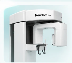
NewTom GO 3D with CEPH, paired with a clinically justified approach to dental imaging.
Our imaging system
NewTom GO 3D with CEPH
One platform for three-dimensional dental imaging, digital panoramic radiography and cephalometric examinations.
10 × 10 cmMaximum documented 3D field of view
80 µmVoxel size in supported high-resolution protocols
2D + 3DPanoramic and volumetric imaging
CEPHCephalometric image acquisition
These are manufacturer-documented capabilities. Field of view, voxel size and exposure options depend on the selected protocol and configuration; they are not universal settings for every scan.
Illustrative visual · Not a photograph of DentoRad equipment
The capabilities, explained
Technology with a clinical purpose.
The useful question is how each feature supports the prescribed examination.
01Coverage
Selectable fields of view
The supplied GO brochure documents selectable 3D volumes up to 10 × 10 cm, including smaller fields for a focused region. Coverage is chosen to include the anatomy needed for the clinical question.
A larger field is not the default for every examination.
02Image sampling
High-resolution protocols
Supported high-resolution protocols can acquire at a voxel size down to 80 µm. The chosen sampling, exposure and reconstruction need to suit the examination.
Voxel size alone does not guarantee diagnostic detail. Movement, metal and noise also matter.
03Exposure adaptation
SafeBeam™
NewTom describes SafeBeam as automatic exposure adaptation to the patient’s anatomical characteristics in the selected area. It supports exposure management within the acquisition workflow.
Automatic adaptation complements clinical justification and protocol selection.
04Protocol choice
ECO acquisition options
ECO protocols are designed to reduce exposure for suitable diagnostic tasks. The required level of detail determines whether an ECO acquisition is appropriate.
ECO, field of view and resolution are protocol-dependent choices, not universal settings.
05Image processing
aMAR
The autoadaptive Metal Artifact Reduction algorithm is designed to reduce artefacts associated with dense materials such as restorations or implants.
Metal artefacts may remain. Processing cannot recover all obscured information or guarantee a diagnosis.
06Panoramic imaging
MultiPAN
MultiPAN produces five panoramic focal layers from one supported acquisition, helping the operator choose the useful layer for the examination.
These are 2D panoramic layers; MultiPAN is not a CBCT volume.
Coverage and detail
Chosen for the question. Considered for the patient.
A focused tooth-related request and a wider jaw assessment may need different acquisition choices.
The supplied GO documentation lists these selectable 3D volumes. The imaging team confirms the appropriate supported combination rather than treating the maximum field and finest voxel as a default.